Photodynamic therapy (PDT) represents an elegant and productive light-based cancer treatment method. This therapeutic modality employs a photosensitizer (PS), which, when activated with light of an optimal wavelength and intensity, elicits a photodynamic reaction, destroying tumors and blood vessels.
The mechanism of action of PDT is based on several key elements. A photosensitizer is a naturally occurring or synthetic substance that transmits light energy when activated by light. An efficacious photosensitizer (PS) should be non-toxic before activation, hydrophilic, activated by a clinically useful light wavelength, and concentrated in tumor tissue rather than healthy tissue. The activation of each PS requires a specific wavelength of light. For instance, red light with a wavelength of 630 nanometres can penetrate tissue to a depth of approximately 0.5 centimetres. The light sources utilized in this process can range from multispectral lamps to precision lasers.
Once the photosensitizer is activated by light, a series of photochemical reactions ensue, destroying the tumor without damaging surrounding healthy tissue. The most significant of these is the Type II reaction, in which the PS, in the presence of oxygen, generates singlet oxygen, which is toxic to cancer cells.
Photodynamic therapy (PDT) is a widely utilized modality in the field of oncology, employed for the treatment of an array of cancers, including those affecting the skin and head and neck regions. Photosensitizers such as hematoporphyrin (HPD), m-tetrahydroxyphenyl chloride (mTHPC), and aminolevulinic acid (ALA) are employed in clinical practice. The mechanisms of action of PDT encompass both cellular and vascular processes. PDT can result in cancer cell death by either apoptosis (programmed cell death) or necrosis (unprogrammed cell death). Additionally, PS can concentrate in vascular endothelial cells, leading to the destruction of the vessels supplying the tumor and hypoxia.
Furthermore, PDT can enhance the immune response, leading to long-term tumor control through the activation of immune cells such as macrophages and T cells.
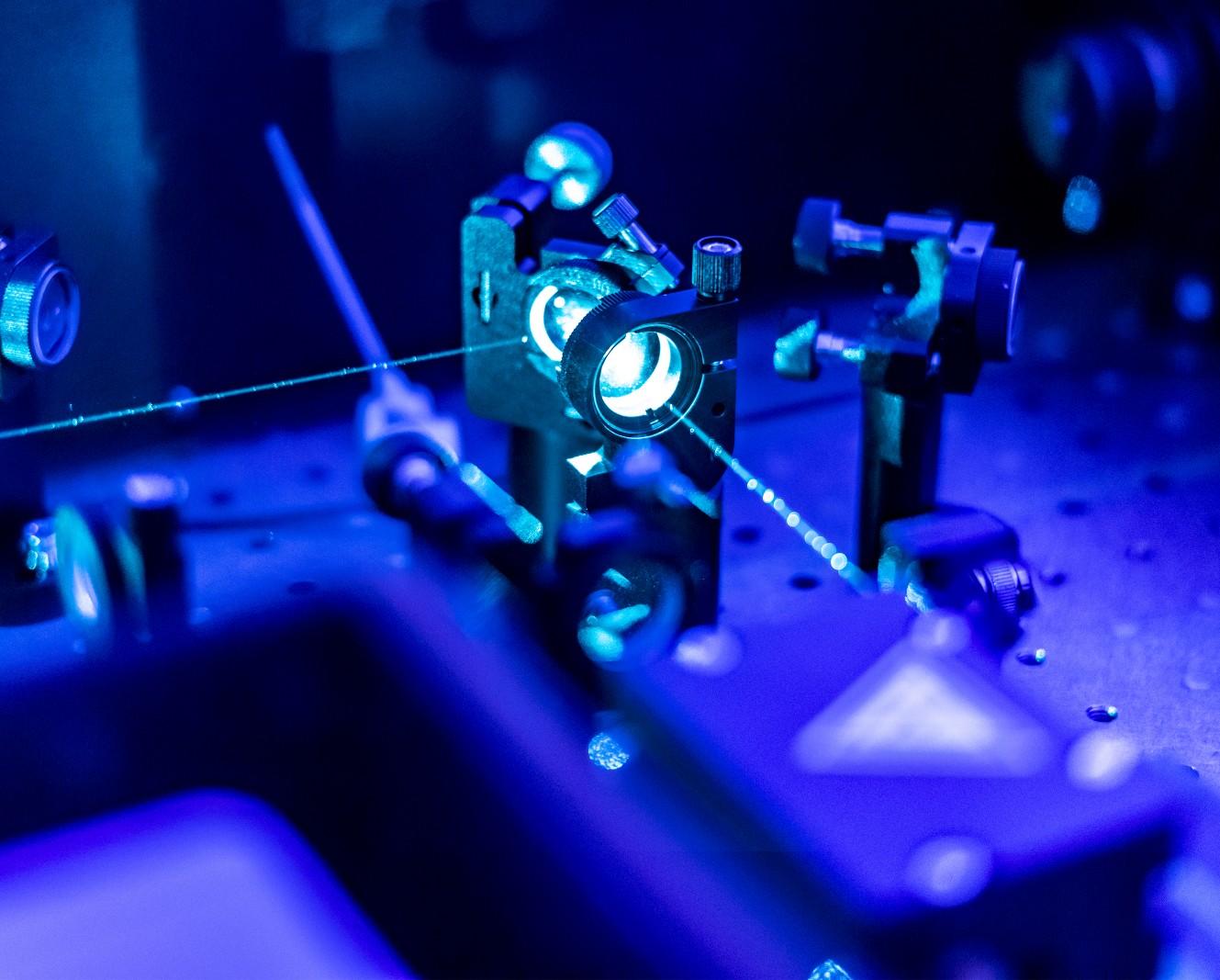
Sonodynamic therapy (SDT) is a modern, non-invasive therapeutic method that uses ultrasound to activate special chemical compounds called sonosensitizers (SS). Under the influence of ultrasound, sonosensitizers enter an excited state, leading to the generation of reactive oxygen species (ROS). These highly reactive molecules can destroy harmful cells, such as cancer cells or bacteria, with minimal impact on healthy tissues. SDT combines mechanical, thermal, and cavitational effects, making it a versatile therapeutic tool.
The mechanism of SDT involves the accumulation of the sonosensitizer at the target site, such as a tumor or bacterial infection. Upon exposure to low-intensity ultrasound, the sonosensitizer becomes excited and releases energy, leading to the formation of ROS. These molecules induce oxidative stress in cells, damaging cell membranes, organelles, and key biomolecules such as proteins and DNA, ultimately leading to cell death. An important feature of ROS is their selective action, allowing SDT to be used in hard-to-reach areas or those requiring precise treatment.
Applications of SDT are broad and primarily include cancer therapy and the treatment of bacterial infections. In oncology, this method is particularly effective for solid tumors, including those located deep within tissues. Ultrasound can penetrate up to 10 cm, making SDT a much more versatile tool compared to photodynamic therapy (PDT), which is limited to surface areas due to light penetration constraints. Moreover, SDT can be used in patients with cancers resistant to traditional treatments, offering new therapeutic possibilities. In treating bacterial infections, SDT proves effective against antibiotic-resistant bacteria and in eliminating bacterial biofilms, which are difficult to remove with standard methods.
The advantage of SDT therapy also lies in minimizing side effects compared to traditional methods such as chemotherapy. By precisely delivering ultrasound to a specific area, damage to surrounding healthy tissues can be avoided. Additionally, SDT can be combined with other treatment methods, enhancing therapy effectiveness. For example, combining SDT with antibiotic therapy or immunotherapy yields synergistic effects, making this method particularly promising for complex clinical cases.
As a next-generation therapy, SDT has enormous development potential. Research is ongoing to improve sonosensitizers, enhance their targeting to specific tissues, and increase ROS generation efficiency. The introduction of carriers such as liposomes or nanoparticles allows for more precise delivery of active substances, further increasing the effectiveness and safety of the therapy. With its unique properties, SDT is becoming one of the most promising tools in cancer and infectious disease medicine, offering new treatment perspectives in an era of increasing antibiotic resistance and the need for less invasive therapies.


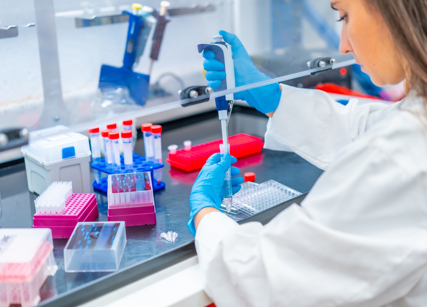
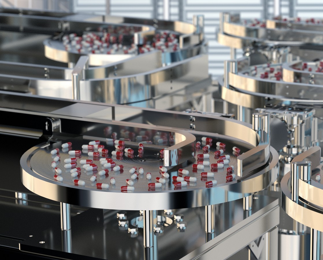
The pharmaceutical pipeline drives the need to develop new oral dosage forms. Our research aims to design solid oral dosage forms by using phospholipids in the form of the (co-)amorphous solid dispersions of poorly soluble drugs using solvent (freeze-drying) and solvent-free (HME) methods. The biopharmaceutical evaluation focuses on cell-free in-vitro tools to assess the dissolution and dissolution/permeation performance. The research focuses on reducing animal use by discovering the underlying mechanism governing the enhanced bioavailability of supersaturating formulations like ASDs.
Biomolecular corona is the sum of biomolecules, which are adsorbed on the surface of nanoparticles (NP) when they contact a biological environment. After corona formation, nanoparticle−corona complexes must be separated from unbound biomolecules and recovered before further analysis. The separation step is crucial and challenging for any studies related to nanoparticle-corona complexes, such as characterization or biological role evaluation. To address the challenges above, we aim to improve the analytical methodology required to understand better the role of biomolecular corona in nanoparticle-mediated drug delivery.
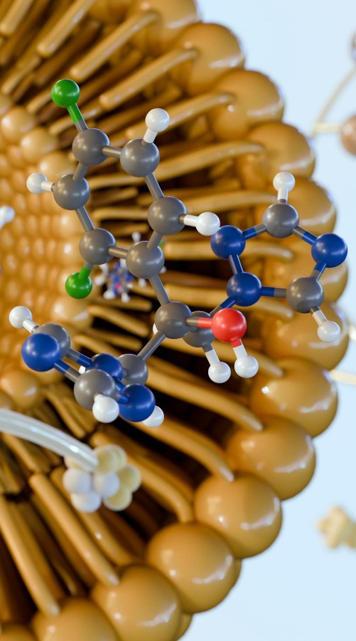
Liposomes and lipid nanoparticles have been proven as useful drug carriers, mostly for anticancer and antifungal drug substances, as well as nucleic acid. In particular, RNA therapeutics have gained significant interest in recent years.
The structure of liposomes and lipid nanoparticles can be modified in many different ways. Also, active ingredients can be embedded in those nanoparticles in various compartments, i.e., encapsulated in the inner aqueous core, incorporated in a phospholipid membrane, or covalently attached to the surface. Our research focuses on the development of dual-loaded or multi-loaded nanoparticles that deliver cargo and substances possessing various functions.